Our practice is committed to empowering our patients with the knowledge and confidence necessary for making informed decisions about their dental health. We strive to offer the highest standard of endodontic care, ensuring that you are well-informed and comfortable with every aspect of your treatment plan.
What to Expect
While each patient’s procedure may differ somewhat, your root canal will generally involve a first meeting to diagnose the problem. The actual procedure may start during that appointment or be scheduled as soon afterwards as possible. Sessions usually last 1.5 hours, and most procedures take 1-2 sessions.
We want to make your dental care more accessible by navigating dental insurance intricacies, offering payment options, and assisting you with any questions regarding financial policies or financing options.
We accept several insurance company plans - please contact us with any questions about coverage.

Our Team
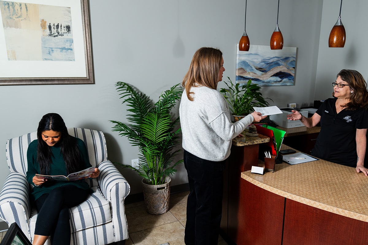
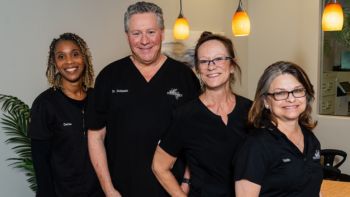
Our caring and compassionate staff begins at our front desk. Kristin has had years of experience as a patient liaison and managing a front desk, and is well versed in handling your insurance needs. Our assistants, Denise and Marcy, are highly skilled and trained in digital technology as well as 2D & 3D imaging. They are very knowledgeable to answer any questions regarding your unique situation. This amount of experience is critical in achieving a smooth and stressless appointment for our patients.
Finance and Pay with CareCredit
CareCredit® is available to help you pay for dental procedures your insurance doesn’t cover. They offer low minimum monthly payment options so you can take care of your dental health. Please click here to apply and pay your through CareCredit.
Pay Your Bill Online
For your convenience, we offer online payment options. Please click here to pay your balance online.
Looking for a practice in the